
MRI has revolutionised the way we diagnose and manage cancer and abdominal diseases. MRI allows us to visualise the internal structures of the body with high clarity, providing detailed information about the location, size, and characteristics of lesions and tissues. With this information, physicians can develop more accurate treatment plans tailored to each patient’s specific needs.
Quantitative MRI
No two patients are alike. Therefore, patients can benefit greatly from personalised treatments. Quantitative MRI (qMRI) biomarkers can characterise tissue diffusion (diffusion-weighted imaging; DWI/intra-voxel incoherent motion; IVIM), perfusion (dynamic contrast-enhanced; DCE/IVIM), hypoxia (Bold/T2*-relaxometry), and elastic properties (magnetic resonance elastography; MRE), four key characteristics that can be used to select the optimal personalised treatment.
Oncology
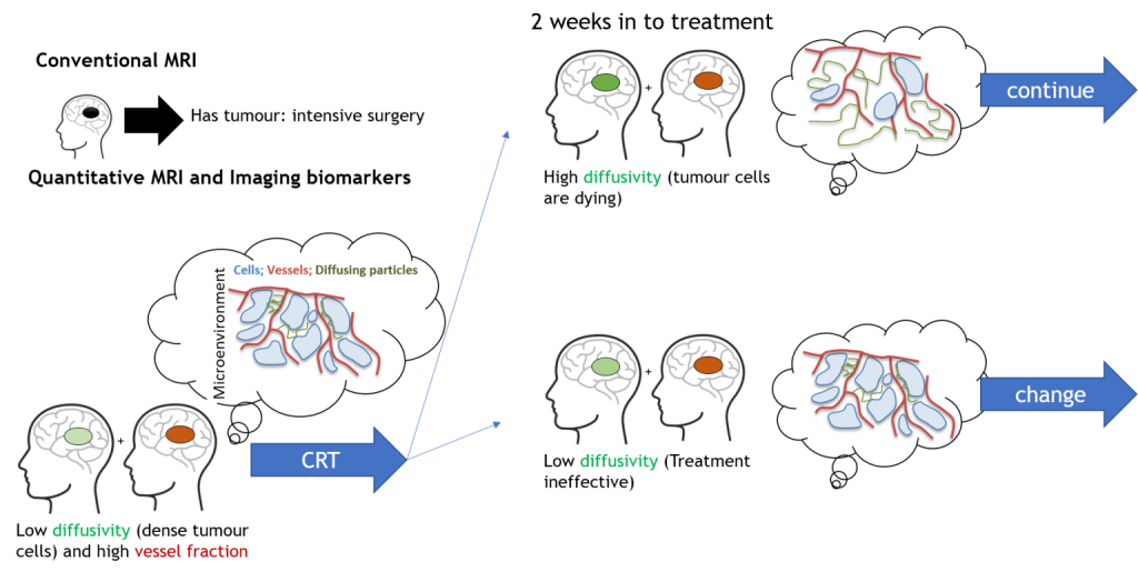
For example, in oncology, good blood flow is essential for chemotherapy and radiotherapy, hence, by characterising blood flow, it will be possible to predict whether these treatments will work even before radiation and chemotherapy. Tumours often have a higher cell density. So, the response to therapy can be determined by measuring a decrease in cell density over time. Therefore, quantitative MRI could have an important clinical impact and enable rational choices for starting, modifying or continuing the most optimal therapy. Our group has several studies investigating the use of quantitative MRI for these purposes in pancreatic, head-and-neck, oesophagal and cervical cancer. (e.g. https://doi.org/10.1002/1878-0261.12688)
Other abdominal diseases
Quantitative MRI may be used to characterise tissue status. For example, investigate the use qMRI to characterise and predict disease progression in metabolic dysfunction-associated fatty liver disease (MAFLD), where we not only measure fat content, but also perfusion, elastography and diffusion. We find that both diffusion and elastography allow distinguishing between reversible simple steatosis and irreversible steatohepatitis. (e.g. https://doi.org/10.1002/jmri.27703)
Quantitative MRI may assess kidney health in patients with Type-2 diabetes. Chronic kidney disease is common in patients with type-2 diabetes. We are investigating the use of quantitative MRI to characterize chronic kidney disease, and see how different treatments affect the kidneys at a functional level.
Patients with abdominal aortic aneurysms can suffer from sudden rupture, which is commonly lethal. Current guidelines to assess the probability of aneurysm rupture and select patients for surgery is solely based on the aneurysm diameter. Our group is currently investigating whether tissue properties, as assessed by quantitative MRI, can help predict the occurrence of aneurysm rupture and highlight patients who benefit from surgery.
Amsterdam UMC
At Amsterdam UMC, we develop and test new quantitative MRI approaches. Notably, we focus on several key aspects. We develop machine learning approaches for improving the quality of quantitative MRI data. Moreover, we develop new techniques to obtain data in the presence of (respiratory) motion. Finally, we test these techniques in clinical studies.
Current projects
- VIDI: Self-supervised physics-informed deep learning quantitative MRI of tumour microstructural imaging for personalized cancer treatments
- KWF-YIG: TOP-QMRI: TOwards Personalized multi-modality oesophageal cancer treatment using machine learning-based Quantitative MRI
- CCA: Non-invasive, accessible liver function assessment with quantitative MRI to guide hepatectomy in hepatic cancer
- Hanarth: The integration of deep learning for quantitative MRI in clinical and research workflows to personalize treatment of cervical and head and neck cancer
- NWO Small NWA: Minimally invasive techniques in oncology: the next steps
- CCA: ROTATION: High Resolution, respiraOry moTion compensATed, abdomInal tumOur imagiNg: Quantitative biomarkers and anatomical imaging
- TKI-PPP: Gamechanger: Development of MRI based endovasculAR procedures for Vascular surgerY (MARVY)
News
Selected Publications
- Gurney-Champion, O. J., Mahmood, … & Redalen, K. R. (2020). Quantitative imaging for radiotherapy purposes. Radiotherapy and Oncology, 146, 66-75.
- Gurney-Champion, O. J., Klaassen, R., … & Nederveen, A. J. (2018). Comparison of six fit algorithms for the intra-voxel incoherent motion model of diffusion-weighted magnetic resonance imaging data of pancreatic cancer patients. PloS one, 13(4), e0194590.
- Kaandorp, M. P., Barbieri, … & Gurney‐Champion, O. J. (2021). Improved unsupervised physics‐informed deep learning for intravoxel incoherent motion modeling and evaluation in pancreatic cancer patients. Magnetic resonance in medicine, 86(4), 2250-2265.
- Ottens, T., Barbieri, S., … & Gurney-Champion, O. J. (2022). Deep learning DCE-MRI parameter estimation: Application in pancreatic cancer. Medical image analysis, 80, 102512.
- Barbieri, S., Gurney‐Champion, O. J., Klaassen, R., & Thoeny, H. C. (2020). Deep learning how to fit an intravoxel incoherent motion model to diffusion‐weighted MRI. Magnetic resonance in medicine, 83(1), 312-321.